The digestive system is where your body breaks down the food you eat into nutrients that can be absorbed into your body.
The digestive system is a series of hollow organs which form a continuous link from your mouth to your anus. These organs include: the oesophagus; stomach; small intestine (duodenum, jejunum and ileum); and the large intestine (colon, rectum and anus). Other organs that are part of the digestive system are the liver, gallbladder and pancreas. In addition, your digestive system is host to a large number of bacteria, known collectively as the gut microbiome, which helps you to digest some foods.
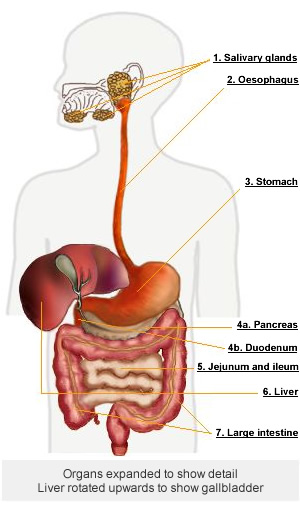
Find out about digestion of your food by viewing the diagram below for each part of your digestive system.

1. Salivary glands
Digestion starts in the mouth, where salivary glands secrete saliva. Saliva is an alkaline fluid which softens food, moistens the mouth and helps swallowing. Saliva also contains an enzyme called amylase, which starts the breakdown of carbohydrate in the mouth.
2. Oesophagus
Food in the mouth is swallowed and then pushed through the oesophagus by muscular contractions and relaxations (called peristalsis) until it reaches the lower oesophageal sphincter, which is a valve that controls food moving from the oesophagus into the stomach and stops it from refluxing back up the oesophagus.
3. Stomach
The stomach is a holding tank with very strong muscular walls. These muscles contract which moves food around and mixes it up.
The stomach lining secretes acidic gastric juices and enzymes to digest carbohydrate and protein. Then the semi-digested food (called chyme) is delivered to the duodenum – the first part of the small intestine – by passing through another valve, the pyloric sphincter.
4a. Pancreas
The pancreas is a tadpole-shaped organ, around 25 cm long, situated behind the stomach in the abdomen. The pancreas produces pancreatic juice which contains enzymes to digest protein, starch and fat. The pancreatic juice is secreted into the duodenum through the duodenal papilla (sometimes called the ampulla of Vater).

The pancreas is also responsible for producing insulin, a hormone which controls blood sugar.
4b. Duodenum
In the duodenum, the chyme, the pancreatic juice and bile from the liver are mixed. The acidic chyme from the stomach is neutralised by the alkaline environment of the duodenum. Food can be digested here in only small amounts, so it’s released from the stomach gradually when there is capacity to process it.
Pancreatic enzymes and bile finish off the chemical breakdown of the acidic stomach contents.
Bile is a greenish-yellow fluid that is made in the liver. Its function is to help in the digestion of fats. It does this by emulsifying fats and breaking down larger globules of fat into smaller droplets, which can be more easily digested.
Bile is stored and concentrated in the gallbladder, a small pear-shaped sac located on the under-surface of the liver on the right-hand side of your abdomen. When fatty foods are consumed, the gallbladder is triggered to release bile into the small intestine via the common bile duct.
If a person has had their gallbladder removed (called a cholecystectomy) bile will drain continuously into the small intestine – and it will become more diluted – as there is no gallbladder to concentrate it. This affects a person’s ability to digest fats – they may have trouble digesting fatty foods. So eating fat after gallbladder removal may need to be managed carefully for a while to avoid discomfort, flatulence (wind) and diarrhoea.
The biliary tract refers to the system of ducts that transport bile from the liver to the gallbladder and small intestine. If a bile duct becomes obstructed (blocked), a person will develop jaundice.
5. Jejunum and ileum
From the duodenum, the mixture is passed into the next section of the small intestine, called the jejunum, and then on to the ileum.
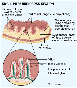
The inside surface area of the jejunum and the ileum is increased by folds and also by millions of finger-like projections called villi and microvilli (see diagram). The villi increase the surface area of the inside of the jejunum and ileum, creating a greater area for absorption of nutrients.

Digestion of foods is completed in this section of the small intestine, and the fats and other nutrients, such as glucose and amino acids are absorbed here through the intestinal wall into the bloodstream and then carried to the liver.
The jejunum has a larger surface area than the ileum, and about 90% of digestion and absorption occurs here, however, the ileum can take on a larger role if the jejunum is damaged or removed. The ileum is the site for absorption of vitamin B12.
Any undigested food, such as fibre, is passed through the ileo-caecal valve to the large intestine.
6. Liver and gallbladder
The incoming blood from the small intestine brings nutrients to the liver for processing, for example, glucose from the breakdown of food is brought to the liver and stored there as glycogen. Other nutrients brought to the liver include amino acids and glycerol.
As mentioned earlier, one of the liver’s functions is to secrete bile, which is then stored and concentrated in the gallbladder.
7. Large intestine (colon)
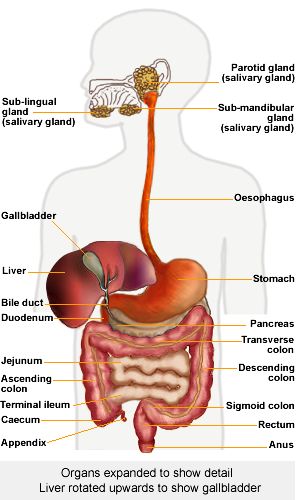
Once the nutrients have been absorbed into the body from the small intestine, the remaining waste is passed into the large intestine (also known as the colon). The large intestine is made up of the caecum, ascending colon, transverse colon, descending colon and sigmoid colon and rectum. The appendix is attached to the caecum.
Content from the small intestine comes into the large intestine as fluid and gradually becomes solid as water and salts are absorbed out as it travels through the large intestine by muscular waves called peristalsis Mucus is secreted to aid the passage of faeces to the rectum.
Stool, or faeces, are stored in the sigmoid colon until a bowel movement pushes them into the rectum and out of the body.
Microbiome
Healthy human beings have roughly as many micro-organisms, such as bacteria, living in and on them as they do cells in their body (trillions). The gut microbiome is the varied population of micro-organisms that live in your digestive system, lining the inside of your intestines. The highest concentration of micro-organisms in the human gut is in the large intestine. Your microbiome is composed of many different species of bacteria. The more varied the species in your population, the healthier it is. The health of your gut microbiome is reflected in your general health.
Some of these bacteria play an important role in digestion, harvesting energy from foods we can’t digest. For example, some gut bacteria can bring about fermentation of undigested dietary fibre in the large intestine. The fibre has made it through our digestive system that far without being digested, because as humans we lack the enzymes to digest it. But the bacteria have the necessary enzymes, fermenting the fibre to produce short-chain fatty acids, compounds which have a role in preventing some diseases and improving the gut barrier.
The bacteria of the large intestine can also synthesise vitamins that humans cannot produce themselves.
The digestive system in more detail
The diagram below shows identifies more of the organs of the digestive system.

Related
ncG1vNJzZmilqZm%2Fb6%2FOpmWarV%2BcrrTA0aigp6yVqMGqusClZKGdkaHBqXvHqK5msZ%2Bqv26yzqibZqGjYrGqs8Ssq56cXw%3D%3D